Structural Anatomy of TIM
To manipulate the molecule just click on it with your left mouse button. Hold the button in while you move your mouse in all directions this should allow you to rotate the molecule in 360 degrees and in 3-D. Also while pressing the shift key move the mouse up or down to zoom in or out. To translate along either the x or y axis hold down both the control key and the right mouse button and move the mouse. To change the molecule into space fill form click here . This shows you the Van der Waals radii of all the atoms in triose phosphate isomerase.
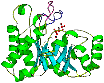
Click here to see a cartoon version of TIM
Click here to see bound substrate (phosphoglycolic acid or PGA an analog of DHAP)
Click here to see only substrate (PGA)
Click here to see hinged lid
Click here to see alternate view of hinged lid
TIM is a homodimeric enzyme comprised of approximately 250 amino acid residues. One complete dimmer is approximately 53,000 Da (i). The structure motif for each sub unit is an eight fold repeat of alpha-loop-beta, with eight parallel beta-sheets in the center surrounded by eight alpha-helixes (iv). TIM dose not contain any metal ion or cofactor. Also there are no alosteric conformations or any cooperation between the two sub units (i). However it has been shown that a monomer of TIM will not catalyze the interconversion (x). The barrel acts as scaffolding to support the active site and substrate while the loops position the substrate and themselves. The active site is located at the C-terminus of one of the barrels. The three main components of the active site are residues his 95, glu 165 and the "hinged lid" (viii). Glu 165 acts as a proton acceptor while his 95 acts as a proton donor (refer to mechanism). There is a "hinged lid" near the active site that clamps the substrate into the active site. This lid is comprised of residues 168 through 174. It moves approximately eight angstroms between its open and closed positions(ix). When the lid is in the closed conformation all solvent is pushed out and the active site is complete. Lid closure is not ligand gated however the entrance of substrate into the active site dose promote lid closure. This is due to the hydrogen bonding of Gly 171 to the phosphate oxygen of the substrate (x).
The following is a diagram of TIM showing both conformations of the flexible loop. The pink colored loop is shown in the open conformation, while the blue loop is in the closed conformation.