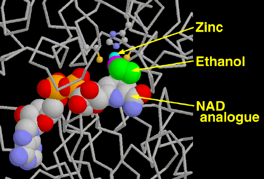
Alcohol dehydrogenase is our primary defense against alcohol, a toxic molecule that depresses the nervous system. High concentrations of alcohol dehydrogenase in the liver and stomach detoxify about approximately one ounce of liquor per hour. The alcohol is converted to acetaldehyde by the oxidation of NAD+ to NADH. Acetaldehyde is even more toxic to the body than ethanol. The body quickly convertes the acetaldehyde into acetate and other molecules that are easily utilized by the cell. Ethanol is not the only target of these enzymes, they also make important modifications to retinol, steroids, and fatty acids.

The NAD Cofactor

Alcohol dehydrogenase uses two molecular tools to convert ethanol to acetaldehyde. The first is a zinc atom which is used to hold and position the alcohol group on ethanol. The second is a large NAD cofactor, which actually performs the reaction. The molecule on the left contains ethanol molecules bound to the two active sites. The ethanol, shown in green and magenta, binds to the zinc and is positioned next to the NAD cofactor. A hydrogen binds to the ethanol carbon and the zinc polarizes the oxygen of the alcohol group. The ethanol carbon loses a hydrogen to NAD and is now acetaldehyde.

Molecular Model of Alcohol Dehydrogenase

Alcohol dehydrogenase is an 80kD protein that functions as a dimer in the cell. It is composed of identical alpha and beta chains.

Alpha and Beta chains

Each chain is composed of 374 residues, composing 12 alpha helices and 19 beta sheets.

Secondary Structure

NAD within the alcohol dehydrogenase subunit

The active site of alcohol dehydrogenase contains a zinc ion that is coordinated to the sulfur atoms of two cysteine residues and a histidine nitrogen atom. The Zn2+ polarizes the carbonyl group of the substrate to stabilize the transition state.

NAD+ Binding Zoom Catalytic Region  

Display entire structure

Use the following commands to adjust the molecular models position. You can also you the mouse to rotate the molecule by clicking on the structure.

Reset Position

Rotate:  Y 90°   Y -90°
  X 90°   X -90°

Distances:  Width (Å)   Height (Å)

Background Color: White Blue Black

NAD Home Introduction Biosynthesis of NAD Example II: Glyceraldehyde-3-Phosphate Dehydrogenase Example III: Lactate Dehydrogenase References