Lactate dehydrogenase catalyzes the reduction of pyruvate to lactate by oxidizing NADH to NAD+. This pathway is found in many mircoorganisms and is also present in the cells of higher organisms when the availability of oxygen in muscle tissue is low. In biochemical terms, lactate is a dead end in metabolism. The reduction of pyruvate to lactate serves only to regenerate NAD+ to allow glycolysis to continue in skeletal muscles. In order for the body to use lactate, it must convert it back into pyruvate through the gluconeogenic pathway in the liver.
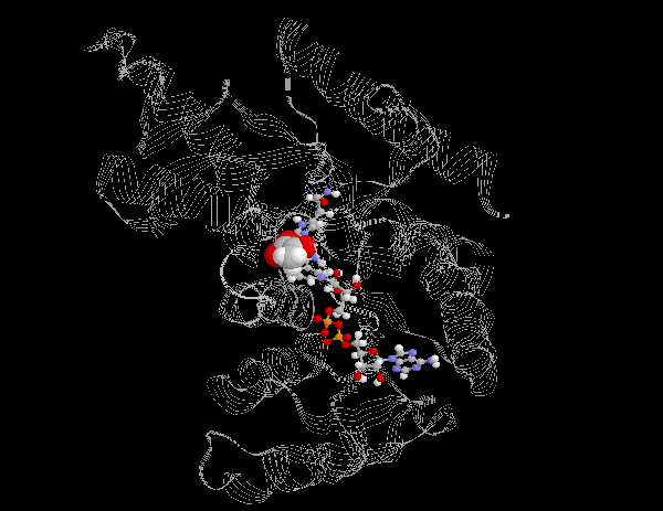
The NAD Cofactor in Lactate Dehydrogenase

Lactate Dehydrogenase is a tetramer composed of two different 35kD subunits, M and H. The H type is found predominantly in the heart while the M type is found primarily in muscle and the liver. The M and H subunits associate to form five different tetramers. These tetramers, called isozymes, can be composed of the following subunit combinations: H4, H3M1, H2M2, H1M3 and M4. The M4 subunit is optimized to convert pyruvate to lactate.

Molecular Model of Lactate Dehydrogenase

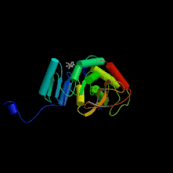
Lactate Dehydrogenase functions as a tetramer. Each subunit is composed of 317 residues, including 10 alpha helices and 15 beta sheets. The structure on left represents one subunit. To view the monomer as a cartoon, press here:

Monomer

To view the secondary structure, press here:

Secondary Structure

To view NAD's position within the enzyme, press here:

NAD within Lactate Dehydrogenase

NADH must bind to lactose dehydrogenase before pyruvate. A hydride ion is transfered, giving a mixture of the two teranary complexes, enzyme-NAD+-lactate and enzyme-NADH-pyruvate. Lactate dissociates from the enzyme followed by NAD+.

NAD+ Binding Zoom Catalytic Region  

Display entire structure

Use the following commands to adjust the molecular models position. You can also you the mouse to rotate the molecule by clicking on the structure.

Reset Position

Rotate:  Y 90°   Y -90°
  X 90°   X -90°

Distances:  Width (Å)   Height (Å)

Background Color: White Blue Black

NAD Home Introduction Biosynthesis of NAD Example I: Alchohol Dehydrogenase Example II: Glyceraldehyde-3-Phosphate Dehydrogenase References