Human  Lysozyme


Structure: Overview

     Human lysozyme (HL) is made up of 130 amino acids and has a macromolecular weight of 14,691daltons, calculated by electrospray ionization mass spectrometry.[Booth et al., 1997]  It is a single strand enzyme with no subunits.  The secondary structures of HL can be broken down into seven helices and one beta sheet consisting of three strands.
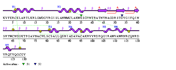
     The seven helices are made up of residues 5-14, 25-35,81-85,90-100,105-108, 110-115, 122-124.[PDB 1LZ1]  Press here to view the seven helices.  Note two of the seven helices are colored blue. Press here to view the helices in the overall model.  The three strands of the beta sheet are made up of residues 43-46, 51-54, and 59-60.  Press here to view the beta sheet.  Press here to see the beta sheet in the overall model
     There are four intramolecular disulfide bonds between cysines which help to maintain the folded structure of HL.  These bonds are between cystine 6 and 128, cystine 30 and 116, cystine 65 and 81, and cystine 77 and 95. ( The first cystine of each pair is colored green and the second is colored yellow.)  To see all four disulfide bonds on one molecule press here.  Below is figure 5 which shows the residue sequence of HL and allows for visualization of where the secondary structures are.


To view a close-up of the protein, position cursor on
image and press the left mouse button +shift key 
and then drag mouse.  To rotate molecule, move the cursor to picture and then press the mouse button and drag the mouse.
Press here to return to a wireframe model.


Figure 5 Sequence of human lysozyme with secondary structure shown. Source: 1LZ1


smuwec.gif (1850 bytes) Home Introduction Reaction Mechanism Active Site Conclusion