The Histone in Action!
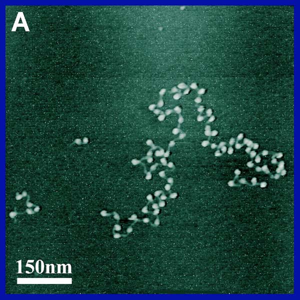
A histone is a protein complex of 5 proteins best known for organizing DNA and coiling it into chromosomes during cell division. Histones are specific to eukaryotic cells only. histones work by forming an octamer. It is considered to be a tetramer of H3-H4 and 2 dimers of H2A and H2B (Gloss). Although in this project, I treat them as all seperate dimers, for ease of showing their individual characteristics. This octamer acts like the wire spool which DNA wraps itsef around. One of the 8 proteins in the middle has a strand which is meant to bind the other protein that connects the lagging DNA to the core histone. Below is a picture of the classic histones electron microscropy shot of "beads on a string." This is the structure seen just before the histones coil and then super coil.
This figure shows the general idea of how histones work. Notice how the beads on a string become a coil, then a supercoil then finally the coil of a coil.
| As you can see to the left are the 5 major DNA configurations.
1. Normal double helix. 2. "Beads on a string" as the DNA winds around the histones. 3. The histone/DNA complexes are beginning to wind up on one another as they form a telephone cord like structure. 4. The supercoiling effect. 5. The coiling of a coil as the DNA enters into the final chromosome structure. |
||||
 |
||||
As you move through this web site you will learn how DNA binds to the histones, where it binds , and how the histone dimers interact with one another!