Structural Characteristics of Lactate Dehydrogenase
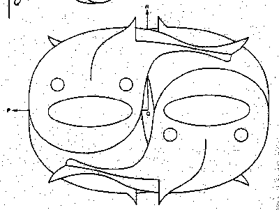
| The picture to the left is of lactate dehydrogenase(LDH). All mammalian cells posses LDH, but the activity of the enzyme varies from tissue to tissue. This variation is due to there being five form of the enzyme called isozymes or isoenzymes. The five isozymes are M4, M3H, M2H2, MH3, and H4. Each isozyme posses a different Km (or affinity) for pyruvate. Each isozyme consists of four subunits of either type M (muscle) or H (heart). While there are some structural differences in the M and H forms, they are similar enough to allow hybirds of the two to form. Illustrated below is the manner in which monomers come together to form a tetramer. |
In subunit-subunit interactions the Q-axis interface dominates and accounts for 50% of the surface area lost by a subunit when a tetramer is formed. These Q-axis contacts consist primarily of hydrophobic interactions between amino acid side chains.
The R-axis contacts are the most polar with 25% of it contact area due to main chain interactions.
The P-axis interface accounts for less than 20% of the lost suface area upon tetramer formation.
The tails of the enzyme subunits are involved in extensive intramolecular contacts with the neighboring subunit.
|
Previous |